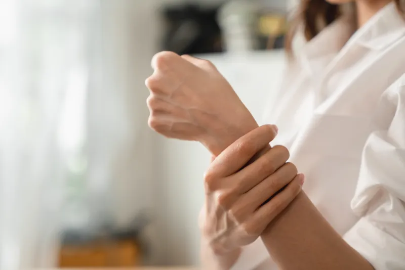
Hypermobility Ehlers-Danlos Syndromes (hEDS)

Hypermobility Ehlers-Danlos Syndromes (hEDS)
Hypermobility Ehlers-Danlos syndromes (hEDS) represents one variant within the broader Ehlers-Danlos syndrome (EDS) classification. Historically known as EDS type III, hEDS is distinguished by generalised joint hypermobility, musculoskeletal manifestations, and certain skin features. Unlike other major types of EDS, the molecular defect causing hEDS remains unidentified (Tinkle et al., 2017).
Epidemiology
- A study involving 12,853 participants found that 3.4% demonstrated joint hypermobility and widespread pain, indicators suggesting potential hEDS (Mulvey et al., 2013).
- A twin study evidenced a genetic component: among dizygotic twins, the concordance rate for joint hypermobility stood at 36%. This rate increased to 60% among monozygotic twins (Hakim et al., 2004).
- The gender disparity is noticeable, with female-to-male ratios ranging from 9:1 to 2:1 (Castori et al., 2010). Chronic pain incidents are also higher among females, with hormonal fluctuations and psychological factors believed to play a role.
Clinical Presentation & Progression
Three progressive stages characterize hEDS:
- Hypermobility Phase: Symptoms include fatigue, lower limb pain, pain on fine motor control, and developmental delays such as dyspraxia. Individuals may experience isolated joint issues or more widespread musculoskeletal pain.
- Pain Phase: Ranging from the twenties to forties, this phase has shared characteristics with fibromyalgia and other chronic overlapping pain conditions. Common manifestations include gastrointestinal complaints, chronic fatigue, orthostatic intolerance, anxiety, sleep disturbances, and more (Tinkle et al., 2017).
- Stiffness Phase: This phase is marked by escalated pain and reduced joint mobility.
Skin & Fascial Manifestations
The skin is comparatively less elastic than in other EDS variants. Pelvic and abdominal diaphragm failures (Nelson et al., 2015) and hernias due to fascial weakness (Nazeem et al., 2013) are more frequent.
Neuropathic Involvement in hEDS
Neuropathic pain, often described subjectively, is common in hEDS. One study pinpointed small fiber neuropathy as a potential source (Cazzato et al., 2016). Genetic abnormalities in TNXB and collagens within nerve structures, combined with joint subluxations, might contribute to this (Voermans et al., 2009; Granata et al., 2013).
Diagnosis of hEDS
hEDS exhibits autosomal inheritance but with an unidentified molecular basis. For clinical diagnosis, the simultaneous presence of three criteria is essential:
- Generalized Joint Hypermobility (GJH): Determined through the Beighton score and adjusted by age and gender criteria. Additionally, the Five-Point Questionnaire, adapted from Grahame and Hakim (2003), aids in assessing GJH.
- Presence of Specific Features: At least two out of three features (A-C) need to be present, encompassing systemic manifestations of connective tissue disorders, a positive family history, and musculoskeletal complications.
- Essential Prerequisites: Absence of unusual skin fragility, exclusion of other heritable and acquired connective tissue disorders, and exclusion of alternative diagnoses.
Management
Due to their susceptibility to small nerve fibre neuropathy, hEDS patients are advised to be cautious about their consumption of fat, alcohol, sugar, and gluten. Limiting these foods can help prevent transient reactions. Anxiety, a well-acknowledged symptom, requires proper support structures. Tailored exercise programs have shown promise, with Swiss ball exercises, in particular, yielding favorable results for improving joint stability.
Testimonial
Thank you, Kieran, for helping me work out how to manage my hEDS and the pain that comes with it. Would struggle without your support. Still learning, but we’re getting there!
Success stories from our clients
EXCELLENT25 reviews on Lesley BeckerKieran is excellent. I cannot thank him enough for his help. Kieran’s professional skills and genuinely kind manner are rare.
 N OKieran has recently provided treatment for my exercise-induced neck pain and migraines. I am a medical doctor. When I was a second-year medical student, I had a head injury that unfortunately led to persistent post-concussion symptoms. I suffered from intractable headaches, migraines, visual disturbances, sensory overload, neck pain, insomnia and exercise intolerance for years. After completing medical school and undergoing specialised post-graduate medical training, I found myself extensively relying on pain relief medications. However, over time, I began to realize that I run out of options. I further trained in functional and integrative medicine, nutrition and acupuncture in hopes of finding some relief. However, my post-concussion symptoms persisted despite all my efforts. Eventually, I came across hyperbaric oxygen therapy ( HBOT ) and had comprehensive training in HBOT. Having found no hyperbaric clinic with the medical standards I was looking for in London, I ended up founding NUMA, my own hyperbaric clinic. HBOT tremendously helped with my spontaneous chronic headaches and some other TBI-related symptoms, but, I still suffered from upper body exercise-induced migraines and intermittent neck pains. If I did not exercise, I was no longer having any migraines. But, I love exercising, and as a medical doctor, I know how important to exercise regularly. I tried physiotherapy to resolve my remaining symptoms which sadly did not help. Eventually, I accepted that I would not be able to do any strength training, and kept walking and doing other forms of exercise that did not involve my upper body. After seeing Kieran helping some of my patients with very complex neurological backgrounds, I decided to have a course of treatment for myself with him. Kieran assessed my condition thoroughly and created a personalised treatment plan for me. He gradually got me back to exercise that involves my upper body too, and completely transformed my threshold for exercise-induced neck pains and migraines. I am a still work in progress, but, I am continuously improving my exercise capacity. I no longer suffer from neck pain. I will continue to refer my patients to Kieran and have my top-up treatments with him. Thank you, Kieran. I could not be more grateful. Dr Nur Ozyilmaz Medical Director of NUMA, Hyperbaric Oxygen Clinic
 ViktorKieran helped me cure my back pain over the long run with only a couple of sessions. His help has allowed me to continue a variety of intensive physical activities pain-free. Top physio.
 Amelia WattsKieran is not only a highly knowledgeable & skilled physiotherapists but also extremely caring. I would highly recommend!
 Agi BorekHighly recommended! I came to Kieran with my knee injury and the treatment he did was very helpful! He also recommended me a few simply exercises I can do at home to recover faster!
 Peter BodiKieran is a very experienced physiotherapist with a wealth of knowledge and practical solutions for the many different manifestation of pain in the lower back.
 jay thakrarI couldn't be more grateful to Kieran for helping me resolve my back pain issues. His thorough and holistic approach was key in reducing my pain levels almost immediately and keeping me free of symptoms long term.
 Maria D'AgostinoI found kieran very competent and knowledgeable in his job. He is very keen on helping and flexible with hours.Load more
